Figure 6.
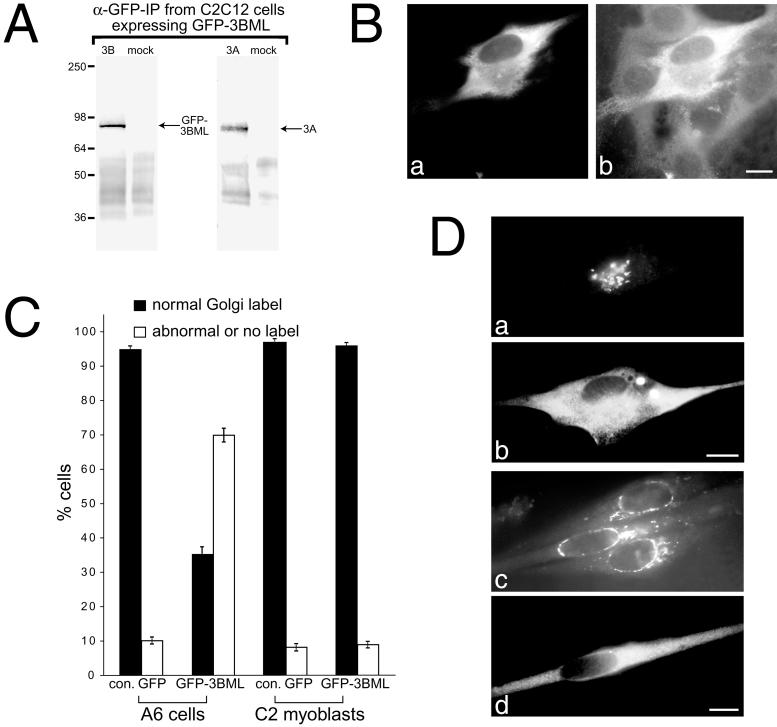
C2C12 myoblasts and myotubes transfected with a KIF3B motorless deletion construct contain normal HP lectin labeling. (A) Motorless-KIF3B dimerizes with endogenous KIF3A in C2C12 cells as demonstrated by the immunoprecipitation of overexpressed GFP-KIF3B-ML from transfected C2C12 myoblasts by using a polyclonal anti-GFP antibody. Anti-GFP coimmunoprecipitates both GPF-KIF3B-ML, as determined by staining with anti-KIF3B-T (to left), and endogenous KIF3A, as determined by staining with the monoclonal K2.4 (to right). Controls were done using Protein A beads alone (mock lanes). (B) Overexpression of motorless-KIF3B induces an increase of endogenous KIF3A expression in C2C12 cells. C2C12 cell transiently transfected with GFP-KIF3B-ML (a) are labeled with the monoclonal K2.4 that recognizes KIF3A (b). Note the increase in KIF3A labeling in the transfected cell compared with the neighboring, nontransfected cells. (C) Quantification of HP lectin labeling of the Golgi apparatus in Xenopus A6 cells and C2C12 myoblasts transiently transfected with motorless-KIF3B (GFP-3BML) and control GFP (con. GFP). Cells (100) were counted for each condition. Bars correspond to the number of cells in each category, and SDs are shown (n = 4 for each condition). Black bars (▪) represent normal HP lectin labeling, whereas white bars (□) represent abnormal labeling or lack of staining altogether. Motorless-KIF3B-transfected A6 cells (60–70%) show abnormal labeling of the Golgi apparatus. This is not seen in motorless-KIF3B-transfected C2C12 myoblasts. (D) Immunofluorescence of motorless-KIF3B in transfected C2C12 cells. GFP fluorescence is seen in a transfected myoblast (b) and transfected myotube (d). The transfected cells are double labeled with Texas Red-conjugated HP lectin (a and c). In the myoblast, HP lectin correctly labels the Golgi apparatus in cells expressing GFP-KIF3B-ML. In differentiating myotubes, the Golgi apparatus becomes redistributed to a ring around the nucleus (c). HP lectin still recognizes the reorganized Golgi apparatus in differentiating cells containing GFP-KIF3B-ML. Bar, 20 μm.

