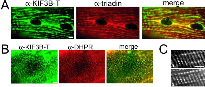
Figure 8.
KIF3B colocalizes with components of the E-C coupling membranes in adult mouse skeletal muscle. (A) Longitudinal section of adult mouse skeletal muscle double labeled with α-KIF3B-T (green) and a monoclonal antibody to triadin (red). (B) Transverse section of adult mouse skeletal muscle double labeled with α-KIF3B-T (green) and a monoclonal antibody to the α-1 subunit of DHPR (red). In A and B, images were colorized in Adobe Photoshop so that areas of overlap in the merged images appear yellow (merge). In A, KIF3B and triadin colocalize in linear points transversely oriented along the surface of the muscle cell. In B, KIF3B and DHPR colocalize within the interfibrillar spaces between the contractile apparatus of the muscle cell. (C) Longitudinal section of adult mouse skeletal muscle double labeled with α-KIF3B-T (a) and a monoclonal antibody to RyR (b). The Z-lines are marked by arrows. KIF3B and RyR are located at either side of the Z-line at the A-I junction. Bar, 20 μm.

