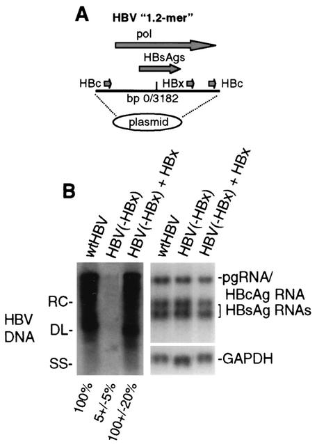
FIG. 1.
HBV replication and transcription in HepG2 cells. (A) Schematic representation of replication-competent HBV genomic DNA. Shown is a head-to-tail replicon of 1.2 copies (1.2-mer) of the HBV genome. Indicated are the duplicated viral core protein (HBc) coding regions, the single HBx coding region, the polymerase (pol) coding region, and the base pair junction of HBV strain ayw. (B) Southern blot analysis of cytoplasmic HBV core particle-associated viral genomic DNA from equal numbers of cells transfected with wild-type HBV, the HBV HBx− mutant, and the mutant trans complemented with an HBx expression vector for 4 days. Replicative DNA intermediates correspond to RC (relaxed circular), DL (double-stranded linear), and SS (single-stranded linear) DNAs. A Northern blot analysis of viral pregenomic (pg), core (HBc), and envelope (HBsAg) mRNAs and cellular GAPDH mRNA obtained from equal numbers of cells in duplicate plates is shown. Quantification of three independent experiments was obtained by densitometry and is shown at the bottom. Data were normalized to those obtained with the wild-type (wt) HBV sample.

