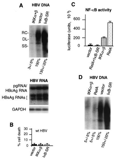
FIG. 5.
Role of NF-κB pathway in suppression of HBV replication. HepG2 cells were transfected with wild-type 1.2-mer HBV genomic DNA and the vector alone or the vector expressing NF-κB-activating kinase IKK-α/β or the NF-κB superrepressor IκB-SR. (A) Southern blot analysis on HBV core particle-associated viral DNA, and Northern blot analysis was performed on poly(A)+ RNA obtained from equal numbers of cells 4 days posttransfection in duplicate plates. RC, relaxed circular; DL, double stranded linear; SS, single stranded linear. (B) HepG2 cells were cotransfected with wild-type (wt) HBV genomic DNA and the vector expressing IKK-α/β or IκB-SR or the vector alone and analyzed for the level of cell death by the LDH assay at 3 days posttransfection. The data shown are averages of three independent experiments. (C) HepG2 cells were transfected with an NF-κB-dependent luciferase transcriptional reporter and the vector alone, the RelA NF-κB subunit expression vector, the vector expressing IKK-α/β, or the vectors expressing RelA and IκB-SR. NF-κB activity was determined by three independent luciferase assays using equal amounts of cellular extracts at 3 days posttransfection. (D) HepG2 cells were transfected with wild-type HBV genomic DNA and the expression vector for IKK-α/β or RelA, the vector alone, or IκB-SR. Viral core particles were isolated from equal numbers of cells at 4 days posttransfection, and viral DNA was assayed by Southern blot analysis. Autoradiograms were quantified by densitometry of at least three independent experiments and normalized to vector-plus-HBV samples.

