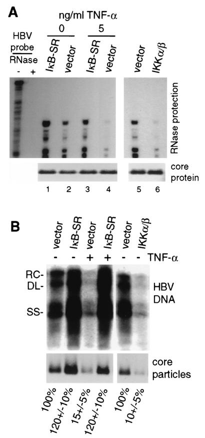
FIG. 6.
Effects of TNF-α and NF-κB on HBV replication, core protein, and particles. (A) HepG2 cells were transfected with wild-type HBV genomic DNA and the vector alone or the expression vector for IKK-α/β or IκB-SR for 4 days. Cytoplasmic core particles were purified from equal numbers of cells, and RNase protection analysis was carried out on the 5′ end of the encapsidated pgRNA. Protected RNA fragments were resolved by denaturing urea-acrylamide gel electrophoresis and autoradiography. Total core protein was determined by SDS-polyacrylamide gel electrophoresis of equal amounts of cellular lysates and detected by immunoblot analysis with antibodies to HBcAg. TNF-α treatment was performed for 1 to 4 days with daily replenishment. (B) HepG2 cells were transfected and left untreated or treated with 5 ng of TNF-α per ml as described above. Southern blot analysis was conducted on HBV DNA extracted from core particles obtained from equal numbers of cells. Core particles were resolved by native agarose gel electrophoresis using the same cell lysates examined for HBV replication and subjected to immunoblot analysis with antibodies to HBcAg. See Materials and Methods for details. Results were quantified by densitometry of at least three independent autoradiograms and normalized to the untreated vector control. RC, relaxed circular; DL, double stranded linear; SS, single stranded linear.

