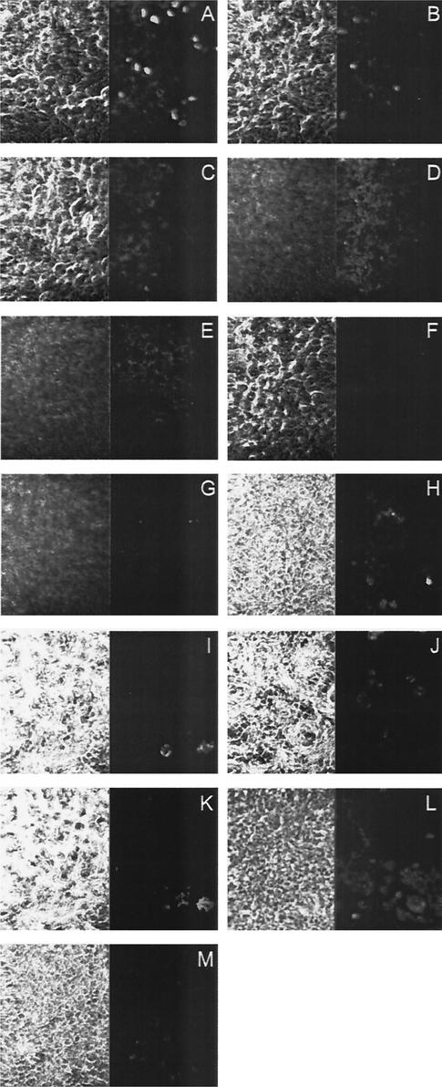
FIG. 4.
Immunofluorescence analysis for transcytosis of 2F5 and 2G12 antibodies. A polarized monolayer of Me-180 cells grown on transwells was incubated with 2F5IgA, human sIgA, 2G12IgM, 2F5IgM, or 2F5IgG from the basal side. After 2 h, the cells were fixed and stained for the human κ light chain. The membrane cell layer was scanned from the basal to the apical side. Pictures A (basal) to E (apical) represent spaced confocal pictures of 2F5IgA-loaded cells. Pictures F and G represent pictures of the basal and the apical side of a cell layer loaded with human sIgA. Pictures H (basal) and I (intra-cellular) show cells loaded with 2G12IgM, pictures J (basal) and K (intracellular) show cells loaded with 2F5IgM, and picture L shows the basal side of cells loaded with 2F5IgG. Picture M shows the negative control (basal) without the addition of antibodies. The left side shows the micrograph, the right side shows the fluorescent signal.

