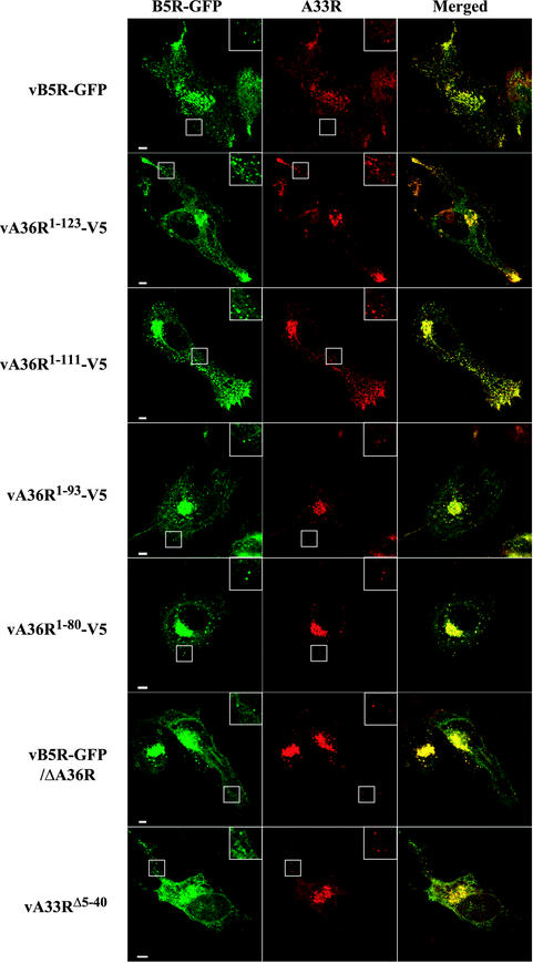
FIG. 4.
Localization of the B5R and A33R proteins in infected cells by confocal microscopy. HeLa cells were infected with the indicated recombinant viruses and were stained with anti-A33R MAb followed by Texas Red-conjugated goat anti-mouse antibody (red). Green fluorescence represents B5R-GFP. Bar, 5 μm. Boxed regions are enlarged to show punctate fluorescence.

