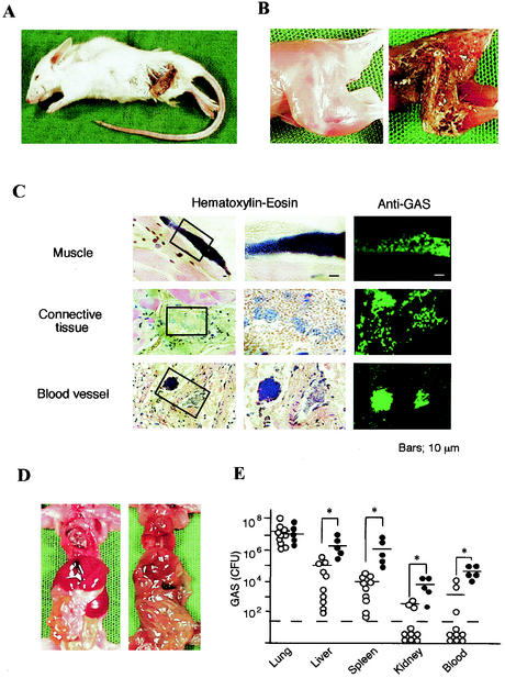
FIG. 3.
Gross pathology of mice with necrotizing fasciitis. (A) Mouse superinfected intranasally with IAV and GAS that died 4 days after the GAS infection. A characteristic appearance of skin necrosis and necrotizing fasciitis can be seen. (B) Hind leg of a noninfected mouse (left) and the dead mouse shown in panel A (right). (C) Histopathology of the necrotic lesion from the mouse shown in panel A. Staining with hematoxylin-eosin (left and middle columns) and immunostaining with rabbit antiserum against GAS and FITC-conjugated anti-rabbit IgG (right column) was done. (D) Internal organs of a noninfected mouse (left) and the superinfected mouse (right). (E) Number of GAS organisms recovered from the lungs, liver, spleen, kidneys, and blood (100 μl) from each dead mouse with (•; n = 5) or without (○; n = 10) necrotizing fasciitis. Open circles under the broken line indicate that any bacteria were not detectable. −, median values; ✽, P < 0.05.

