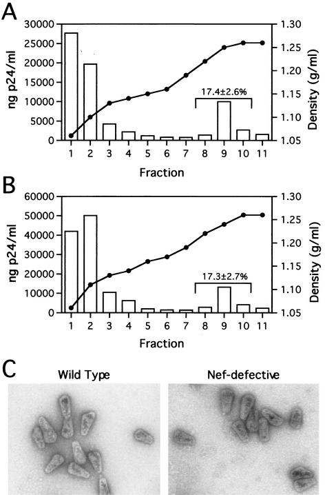
FIG. 1.
Analysis of wild-type and Nef− core yields and morphology. (A and B) Quantitation of wild-type and Nef-defective HIV-1 core yields. Concentrated virions were layered onto 30-to-70% (wt/vol) linear sucrose gradients atop a layer of Triton X-100. Following ultracentrifugation at 100,000 × g for 16 h at 4°C, fractions were collected from the top of the gradient and were analyzed by p24 ELISA. The density of each fraction was determined by refractometry. Panels A and B show the recovery of cores from wild-type and Nef-defective HIV-1 particles, respectively. Values shown represent the mean percentages (and standard deviations) of p24 found in the core-containing fractions for a minimum of five independent determinations. (C) Ultrastructural analysis of wild-type and Nef− cores. Gradient fractions containing intact cores were pelleted at 100,000 × g for 30 min and resuspended in 15 μl of STE buffer. Samples were loaded onto carbon-coated grids and stained with 1% uranyl acetate. Samples were visualized at a magnification of ×40,000 in a Philips CM12 transmission electron microscope.

