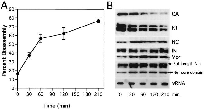
FIG. 3.
Kinetic and biochemical analyses of HIV-1 core disassembly in vitro. Purified HIV-1 cores were incubated at 37°C for the indicated times, followed by separation of soluble and core-associated CA by ultracentrifugation. (A) Dissociation of CA from pelletable cores during incubation at 37°C. Supernatants and pellets were analyzed by p24 ELISA. The extent of disassembly was determined as the percentage of the total CA protein in the reaction mixture detected in the supernatant. Values represent means of triplicate determinations with error bars representing one standard deviation. (B) Biochemical analysis of particles remaining following disassembly of HIV-1 cores. Pellets from disassembly reactions shown in panel A were subjected to Western blotting and RNA slot blot analyses. The Western blot membrane was probed sequentially with rabbit anti-CA, anti-RT, anti-NC, anti-IN, anti-Vpr, and anti-Nef antibodies. RNA was extracted from pellets, denatured, and immobilized by vacuum filtration. Relative HIV-1 RNA levels were determined by hybridization with a 32P-labeled HIV-1 probe, followed by detection with autoradiography.

