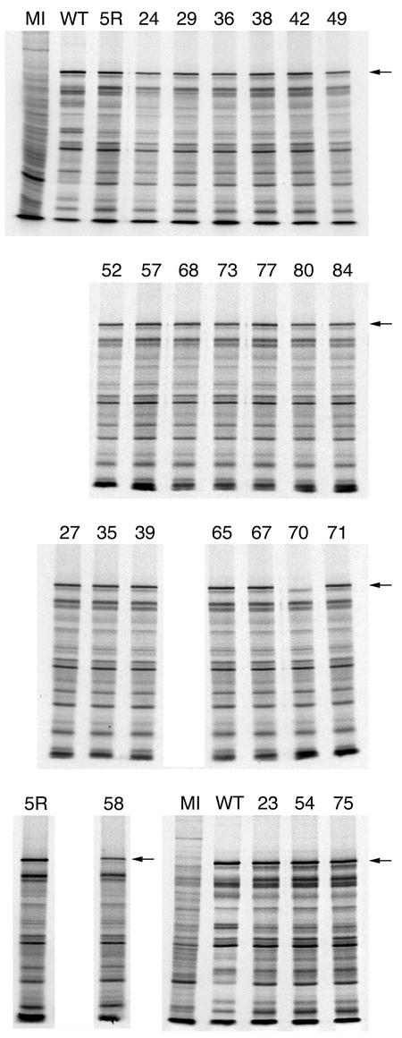
FIG. 4.
Accumulation of ICP5 in the mutant-infected cells. Vero cell monolayers (2.5 × 10⁵ cells) were either mock infected (MI) or infected with wild-type KOS (WT), the marker-rescued virus K5R (5R), or the mutant viruses at a multiplicity of infection of 10 PFU/cell. Infected cells were radiolabeled with [35S]methionine from 9 to 24 h postinfection. The cells were lysed in Laemmli sample buffer, and the proteins were analyzed by SDS-PAGE (9% acrylamide). The autoradiographs obtained following exposure of the dried gels to X-ray film are shown in the figure. The numbers above each lane refer to the mutated residue. The position of the ICP5 polypeptide is shown on the right of the panels (see arrow).

