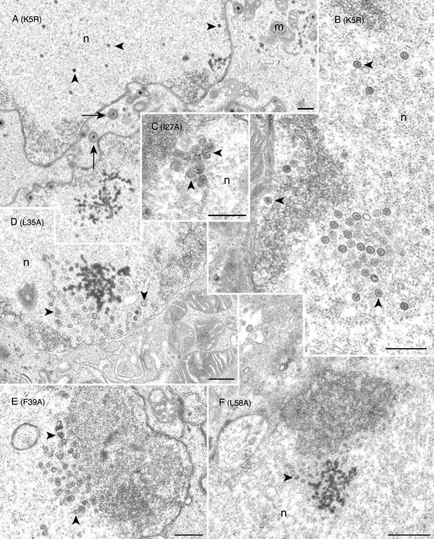
FIG. 5.
Conventional transmission electron microscopy of the mutant-infected cells reveals open shells in the nucleus. Vero cells were infected with K5R (A and B) or with the mutant viruses I27A(C), L35A (D), F39A (E), and L58A (F). Infected cells were harvested 18 h following infection and processed for transmission electron microscopy. Mature capsids (marked by arrowheads) were seen in the nucleus (n), and enveloped virions (marked by arrows) were evident in K5R-infected cells. In the mutant-infected cells (mutation shown in parentheses), numerous large open capsid shell structures (marked by arrowheads) were detected in the nuclei (n). In L58A-infected cell nuclei, smaller capsid shells (see arrowhead) were observed (panel F). Bars, 0.5 μm. Mitochondria (m) are indicated.

