Figure 3.
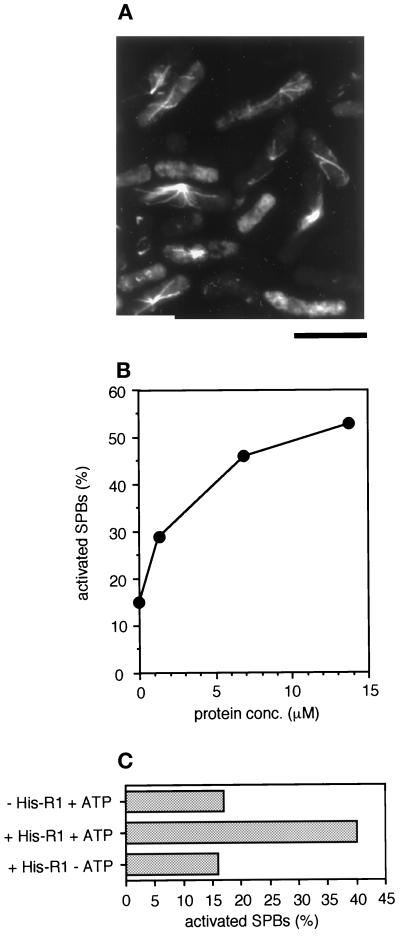
SPB activation by recombinant His-mouse R1 fusion. (A) The SPB activation assay. Permeabilized S. pombe interphase cells were incubated with 59 μM recombinant histidine-tagged mouse R1 (His-R1). Polymerized microtubules were visualized by immunofluorescence with anti–α-tubulin (B-5-1-2). Bar, 10 μm. (B) Protein-concentration dependency of SPB activation by recombinant His-R1 protein. Horizontal axis, the protein concentration of His-R1 during incubation with permeabilized S. pombe interphase cells (N = 300). (C) ATP dependency of SPB activation by recombinant His-R1 protein. Permeabilized S. pombe interphase cells were incubated with a control buffer containing 1 mM Mg-ATP and ATP regeneration system (−His-R1 + ATP) or with 30 μM His-R1 protein in the presence (+His-R1 + ATP) or absence (+His-R1 − ATP) of 1 mM Mg-ATP and ATP regeneration system. Horizontal axis, the percentage of activated SPBs (N = 300).

