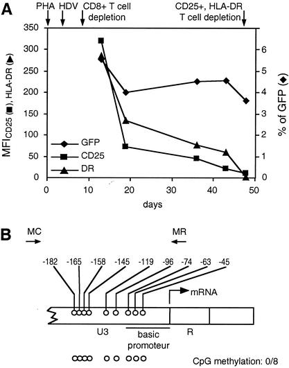
FIG. 4.
Expression of the HDV, cell-surface activation markers, and CpG methylation pattern in the LTR region in primary resting memory T cells challenged in vitro. (A) The PHA-activated PBMC were transduced with HDV, and after the CD8+-T-cell depletion, the percentage of GFP+ cells (⧫) and the MFI for CD25 (▪) and HLA-DR (▴) were monitored for up to 50 days after activation. The time schedule of cell culture and HDV challenge are indicated by arrows. (B) CpG methylation pattern of the LTR region in resting CD4+ T cells at day 50 after PHA activation. Positions and orientations of PCR primers MC and MR used to amplify bisulfite-treated HDV DNA are shown by arrows. The arrow at the U3-R junction denotes the start site of transcription (nucleotide +1). Open circles, nonmethylated CpG residues in 8 tested promoter sequences.

