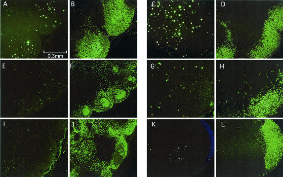
FIG. 5.
SIV Env+ cells in LNs at 2 weeks p.i. Serially sectioned tissue specimens were stained either with an anti-SIV gp32 Env antibody (A, C, E, G, I, and K) or with an anti-CD20 antibody (B, D, F, H, J, and L) to stain B cells. (Left panels) LNs of rhesus macaques infected with Δnef: Mm16 (A and B), Mm14 (E and F), and Mm29 (I and J). (Right panels) LNs of rhesus macaques infected with SIVmac239: Mm18 (C and D), Mm24 (G and H), and Mm27 (K and L).

