
Figure 6.
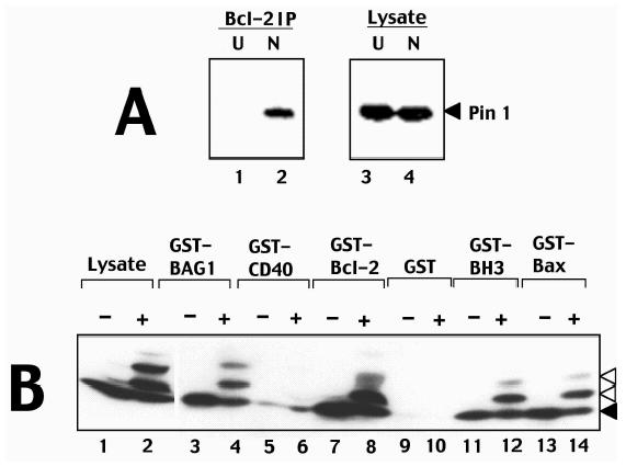
Phosphorylated Bcl-2 interacts with Pin1. (A) Detergent-solubilized lysates (2x10⁶ cells/sample) were prepared either from 293-Bcl-2 cells that were cultured without treatment (lanes 1 and 3) or with nocodazole (lanes 2 and 4). Samples were either analysed directly by immunoblotting with anti-Pin1 antibodies (lanes 3 and 4) or were subjected to immunoprecipitation with anti-Bcl-2 antibodies followed by immunoblotting with anti-Pin1 antibodies. Immunodetection was accomplished by an ECL method. (B) GST-fusion proteins (1 µg) were immobilized on glutathione-Sepharose and incubated with lysates prepared from 697-Bcl-2 cells (100 µg total protein) cultured for ∼1 day with (+) or without (-) 300 nM paclitaxel. Beads were washed extensively and associated proteins were analyzed by SDS-PAGE/immunoblotting using anti-Bcl-2 antibody. An equivalent aliquot of lysate was loaded directly in gels for comparison.