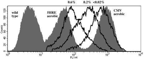
Figure 1.
EGFP fluorescence in HT 1080 human fibrosarcoma cells transfected with the hypoxia-responsive 5HRE-hCMVmp-d2EGFP vector (destabilized EGFP protein, half-life 2 hours), after treatment at oxygen levels between 20% (“5HRE aerobic,” grey area) or the concentrations indicated (black lines) for 12 hours and a subsequent 4-hour reoxygenation period. Fluorescence of positive control cells transfected with CMV-d2EGFP vector (“CMV aerobic,” grey area) and of wild-type HT 1080 cells (grey area) is shown for comparison. Gating for scatter and presence of transfection was performed. Not all oxygen concentrations are shown for clarity. Flow cytometry data are from a representative experiment.

