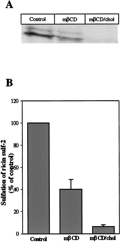
Figure 2.
Effect of mβCD and mβCD/chol on sulfation of ricin sulf-2. (A) Representative example of the resulting bands seen after fluorography in a given experiment. The HeLa-TetOn/Rab9S21N cells were washed with sulfate-free DME medium and incubated for 4 h in the presence of Na235SO4 before addition of 5 mM mβCD or mβCD/chol. After 30 min, ricin sulf-2 was added, and the incubation continued for 2 h. The cells were then washed with 0.1 M lactose in HEPES medium at 37°C and with ice-cold PBS before they were lysed. The nuclei were removed by centrifugation, and the sulfated ricin was immunoprecipitated with rabbit antiricin antibodies attached to protein A-sepharose overnight at 4°C. The immunoprecipitate was analyzed by SDS-PAGE (12%) under reducing conditions followed by fluorography. The intensities of the resulting bands were determined by densiometric quantitation using ImageQuant 5.0. (B) Averaged data of three independent experiments.

