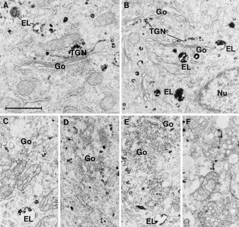
Figure 8.
Electron microscopic pictures of HeLa-TetOn/Rab9S21N cells incubated for 2 h at 37°C with a monovalent conjugate of ricin B-chain covalently coupled to HRP. A and B are from control cells, whereas C-F are from cells treated with 5 mM mβCD/chol for 30 min at 37°C before addition of the ricin B-chain conjugate. In control cells, well-developed Golgi stacks (Go) and TGN labeled by ricin B-chain conjugate are seen. In contrast, following incubation with mβCD/chol, the size of distinct Golgi stacks is strongly reduced and numerous small vesicles accumulate instead. EL, ricin B-chain conjugate labeled endosomes/lysosomes; Nu, nucleus; Bar, 1 m.

