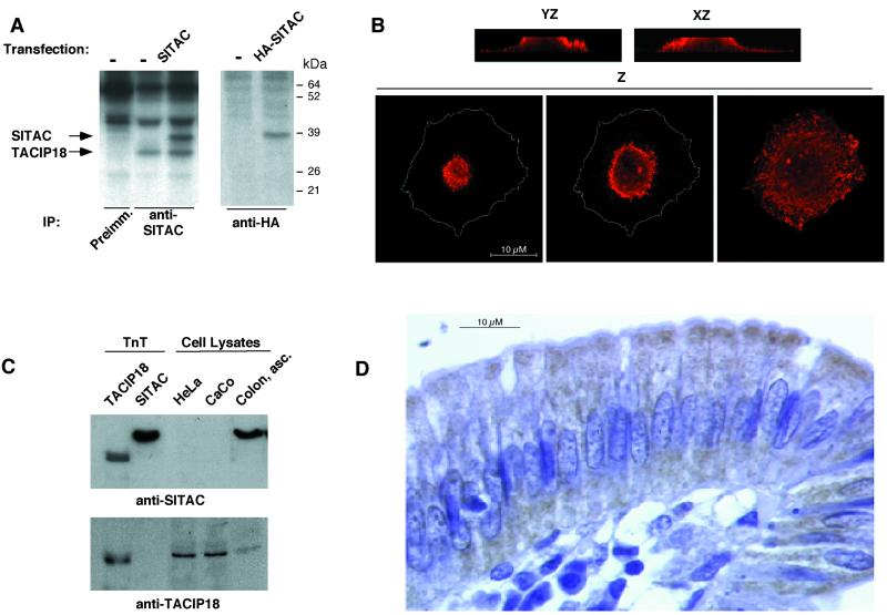
Figure 2.
Expression and subcellular localization of transfected or endogenous SITAC. (A) HeLa cells transiently transfected with SITAC or HA/SITAC were metabolically labeled and immunoprecipitated with anti-SITAC or anti-HA antibodies as indicated. Immunoprecipitates were analyzed by SDS-PAGE and fluorography. (B) CHO transiently transfected with HA/SITAC were permeabilized and incubated with anti-HA antibodies, TRITC-labeled anti-mouse antibodies and analyzed by confocal microscopy. Several consecutive single confocal planes stacked through the YZ and XZ axis, respectively, and three consecutive single confocal planes through the Z axis are shown.The perimeter of the cell as seen in the right panel has been drawn on the left and middle lower panels to show the relative position of the fluorescence within the cell. (C) In vitro transcribed and traslated TACIP18 or SITAC or total cell lysates from HeLa or CaCo cells or from fresh human colon samples were analyzed by western blotting with anti-SITAC or anti-TACIP18 polyclonal antibodies as indicated. (D) Immunohistochemical staining of human intestinal wall with anti-SITAC polyclonal antibodies.

