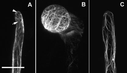
Figure 6.
Effects of CA-rop2 and DN-rop2 Expression on F-Actin Localization in Root Hairs.
F-actin was visualized in live cells using transiently expressed GFP-mTalin as described in the text. Root hairs expressing GFP-mTalin were observed using confocal microscopy. Images shown are projections of scanning laser sections (1 μm) along the axes of root hairs.
(A) In wild-type hairs, axial actin cables end at the subapical region (arrow), whereas the apex contains fine F-actin (arrowhead).
(B) In CA-rop2 hairs, an extensive actin network was found throughout the cortex.
(C) In DN-rop2 hairs, axial actin cables protruded to the extreme apex and no fine F-actin was found in the apex.
For each line, more than six hairs were observed and showed identical or very similar staining patterns. Bar in (A) = 20 μm for (A) to (C).

