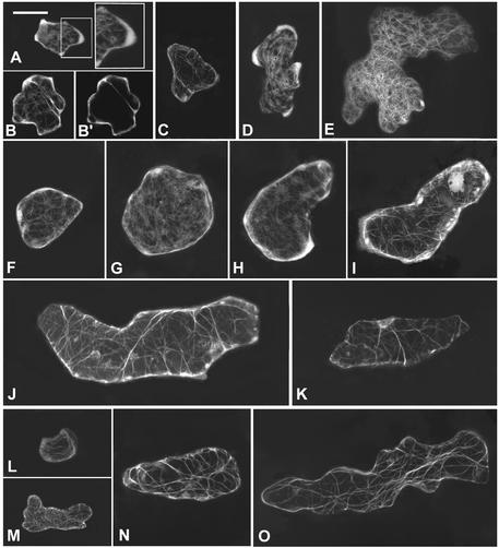
Figure 7.
Formation of Diffuse Cortical F-Actin Is Associated with Cell Expansion and Is Altered by CA-rop2 and DN-rop2 Expression.
To visualize F-actin in live cells, GFP-mTalin was expressed transiently in leaf pavement cells of wild-type, CA-rop2, and DN-rop2 plants, and different stages of cells expressing GFP-mTalin were imaged using confocal microscopy as described in the text. All images shown are projections of serial laser sections except for (B′). Bar in (A) = 25 μm for (A) to (O).
(A) and (B) Wild-type stage I cells contained diffuse F-actin localized throughout the cell cortex.
(B′) Mid-plane section of the cell shown in (B) showing the cortical localization of diffuse actin and a cytoplasmic strand containing F-actin.
(C) A stage I cell showed no diffuse cortical actin when AtPFN1 is coexpressed with GFP-mTalin.
(D) A stage II cell contained strong diffuse F-actin in the cortical region of the lobe primordia.
(E) No diffuse cortical actin was found in a stage III cell, but an extensive network of actin cables was seen.
(F) to (J) CA-rop2 cells showed diffuse cortical actin at all stages: stage I (F), stage II ([G] to [I]), and stage III (J).
(K) Stage II CA-rop2 cell showed no diffuse cortical actin when AtPFN1 is coexpressed with GFP-mTalin.
(L) to (N) DN-rop2 cells contained little diffuse cortical actin in stages I ([L] and [M]) and II (N) and no diffuse cortical actin at stage III (O). Actin cables were present in all stages of pavement cells and were unaffected by CA-rop2 or DN-rop2 expression.

