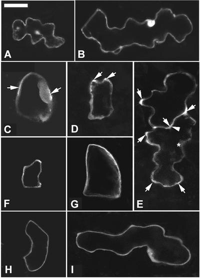
Figure 9.
GFP-Tagged Rop2 Is Localized Preferentially to the Apparently Expanding Region of the PM.
To assess the subcellular localization of ROP2, GFP-ROP2 was expressed transiently in pavement cells and visualized by confocal microscopy. (A) and (B) show cells expressing GFP alone as a control. (C) to (I) show localization of GFP-ROP2. Arrows indicate strong GFP-ROP2 localization to specific cortical regions. Images shown are 1-μm laser sections. Bar in (A) = 20 μm for (A) to (I).
(A) and (B) Stage II and stage III cells, respectively. GFP was localized in the nucleus and throughout the cytoplasm.
(C) and (D) Two wild-type cells at stage I expressing GFP-ROP2.
(E) Two adjacent stage II cells expressing GFP-Rop2. Note a sinus region (arrowhead) in the bottom cell showing weak fluorescence, whereas its opposing lobe region in the neighboring cell showed strong PM fluorescence. Some regions of the cell were rich in cytoplasm, as indicated by the asterisk in the bottom cell, but contained weak fluorescence, indicating that the strong localized fluorescence was not attributable to dense cytoplasm in those regions.
(F) and (G) GFP-ROP2 localization in CA-rop2 pavement cells. GFP-ROP2 was distributed much more evenly throughout the PM in CA-rop2 cells at the equivalent stage (stage I) than in wild-type cells.
(H) and (I) PM localization of GFP-ROP2 in DN-rop2 pavement cells was very weak and was indistinguishable from its cytoplasmic localization.

