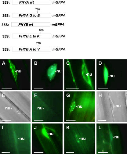
Figure 5.
The Formation of Intranuclear Speckles Is Not Detectable in Transgenic Plants Expressing Mutant phyA:GFP or phyB:GFP Fusion Proteins.
The top shows a diagram of the wild-type (wt) and mutant PHYA:GFP and PHYB:GFP transgenes expressed in Arabidopsis plants. Expression of these transgenes was driven by the viral 35S promoter. The positions of the amino acid substitutions within the mutant phyA and phyB molecules are shown. (A) to (L) show epifluorescence images ([A] to [D], [F], [G], and [I] to [L]) or differential interference contrast images ([E] and [H]) of nuclei in hypocotyl cells in 7-day-old seedlings expressing wild-type phyA:GFP ([A] and [B]) and phyB:GFP ([E] to [G]) and the mutant phyA:GFP ([C] and [D]) and phyB:GFP fusion proteins (position 838 [{H} to {J}] and position 776 [{K} and {L}]) either at the end of dark incubation ([A], [C], [E], [F], [H], [I], and [K]) or after 9 h of FR ([B] and [D]) or 18 h of R ([G], [J], and [L]). (E) and (F) and (H) and (I) are pairs that represent the same cells. Positions of the selected nuclei are indicated (nu). Bars = 10 μm.

