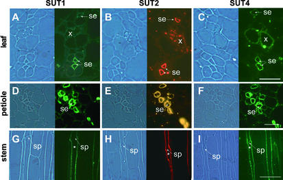
Figure 2.
Colocalization of Different Suc Transporters in Sieve Elements on Serial Sections of Potato Source Leaves, Petioles, and Stems.
(A) to (C) Source leaves.
(D) to (F) Petioles.
(G) to (I) Stems.
SUT1 detection ([A], [D], and [G]) was performed using StSUT1-specific antibodies as described previously (Kühn et al., 1997). SUT2 detection ([B], [E], and [H]) and SUT4 detection ([C], [F], and [I]) were performed using LeSUT2 and LeSUT4 antibodies able to recognize their potato orthologs StSUT2 and StSUT4, respectively (Barker et al., 2000; Weise et al., 2000). SUT1 and SUT4 detection was visualized with a fluorescein isothiocyanate–coupled secondary antiserum, and SUT2 detection was visualized with a cy3-coupled secondary antiserum that was excited either via a broad-spectrum filter set (source leaves and stem longitudinal sections) or via a cy3-specific filter set (petiole cross-sections). The same sieve element in the longitudinal serial section is indicated by asterisks. se, sieve elements; sp, sieve plate; x, xylem. Magnification was ×250. Bars = 20 μm in (A) to (C), 40 μm in (D) to (F), and 100 μm in (G) to (H).

