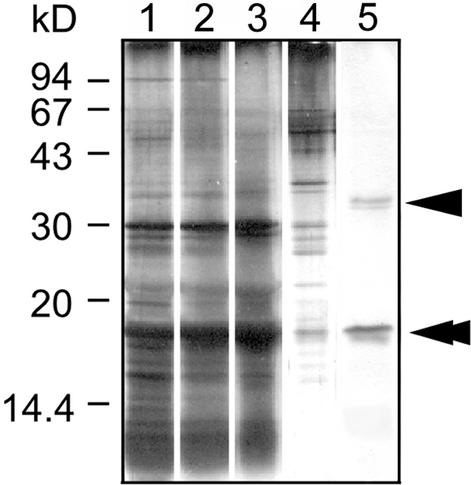
Figure 2.
DNA-Cellulose Chromatography of C. merolae cp-Nucleoid Proteins.
Proteins were analyzed by SDS-PAGE using a 15% gel. Protein bands were visualized by silver staining. Lane 1, isolated cp-nucleoids; lane 2, supernatant of cp-nucleoid subfraction after DNaseI treatment; lane 3, flow-through fraction; lane 4, fraction eluted with 300 mM NaCl; lane 5, fraction eluted with 1 M NaCl. Arrowhead and double arrowhead indicate the 35- and 17-kD proteins, respectively.

