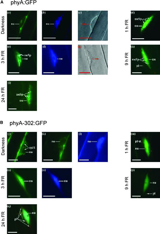
Figure 6.
Arabidopsis phyA:GFP and phyA-302:GFP Fusion Proteins Translocate to the Nucleus, but phyA-302:GFP Fails to Produce Nuclear Speckles under FR.
(A) Representative nuclei of transgenic seedlings expressing phyA:GFP.
(B) Representative nuclei of transgenic seedlings expressing phyA-302:GFP.
The seedlings were kept in darkness ([a], [b], [c], [j], [k], and [l]) or transferred to continuous FR for 1 h ([d] and [m]), 3 h ([e], [f], [g], [n], and [o]), 9 h ([h] and [p]), or 24 h ([i] and [q]). The subcellular localization of the GFP fusion proteins was investigated by fluorescence microscopy ([a], [d], [e], [h], [i], [j], [k], [m], [n], [p], and [q]). 4′,6-Diamidino-2-phenylindole staining ([b], [f], [l], and [o]) and differential interference contrast images ([c] and [g]) are included ([a] to [c], [e] to [g], [k] to [l], and [n] to [o]; each show identical nuclei). Nuclei (nu), nuclear spots (nuSp), plastids (pl), and cytoplasmic fluorescence (cpFl) are indicated. Bars = 10 μm.

