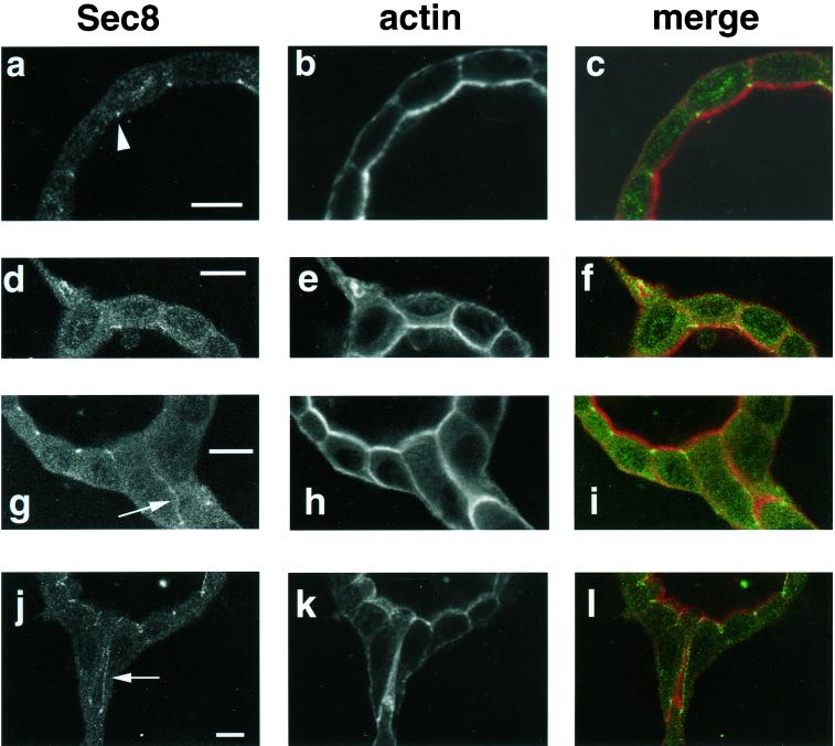
Figure 2.
Sec8p relocalizes during tubulogenesis. (a and b) Fluid-filled cyst formed by MDCK cells grown for 10 d in a collagen gel. Staining is seen at the area of the tight junction (arrowhead) by using anti-Sec8 antibody (a). Sec8p colocalized with the tight junction protein ZO-1 (Figure 1). Concurrent staining of actin with phalloidin was performed (b). (c) Merge of a and b. (d–l) Fluid-filled cysts formed by MDCK cells grown for 10 d in collagen and stimulated for 24 h with conditioned medium containing HGF. The exocyst can be seen relocalizing along the growing tubules in a pattern consistent with the changes in polarity that occur as tubules form. (d–f) Extension stage of tubulogenesis. Sec8p is seen relocalizing into the extension (d), in association with actin staining (e). (g–i) Cord stage of tubulogenesis. Arrow in g indicates staining at the region of cell–cell contact in the cord. This region may become the boundary of a new lumen, as shown by the intense actin staining in this region (h). (j–l) Nascent tubule in the final stage of tubulogenesis. Arrow in j shows two vertical lines of Sec8p staining outlining the boundary of the lumen. Intense actin staining in a broad band in k underlies the apical surface surrounding this lumen. (f, i, and l) Merge of d and e, g and h, and j and k shows that the relocalizing exocyst closely surrounds, but does not precisely overlap the actin in the projections of the nascent tubules as indicated by lack of yellow in the merged panels. Sec 8, green; actin, red. Bar, 10 μm.

