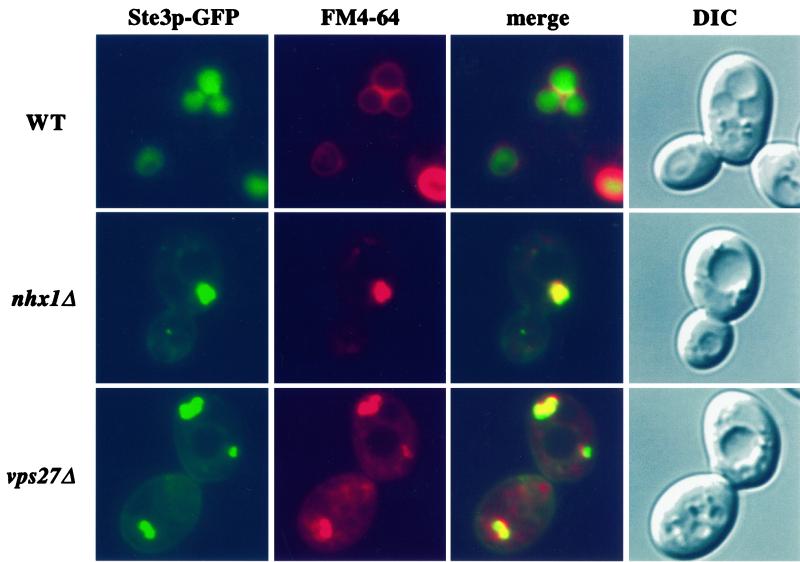
Figure 5.
FM4-64 and Ste3p-GFP accumulate in the class E compartment of nhx1Δ cells. Wild-type (WT; SF838-9Dα), nhx1Δ (KEBY15), and vps27Δ cells (SGY73) were transformed with pJLU34. Cells were labeled in 40 μM FM4-64 for 15 min at 30°C and then chased in fresh medium for 30 min at 30°C. FM4-64 and Ste3p-GFP were photographed under the red and green fluorescence channels, respectively, and a merged image of these two channels is also shown. Differential interference contrast (DIC) images of the same cells were collected to visualize the vacuoles.

