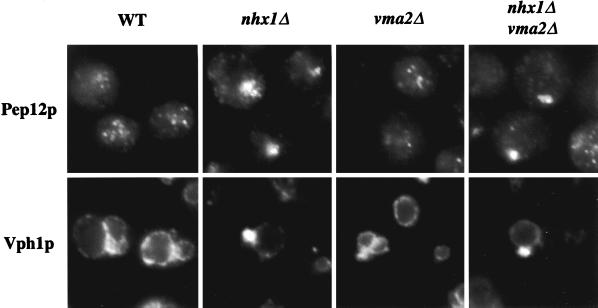
Figure 8.
Unlike nhx1Δ or nhx1Δ vma2Δ cells, vma2Δ cells do not show a class E Vps− morphological phenotype. Immunofluorescence was performed as described in Figure 4, with wild-type (WT: SF8389Dα), nhx1Δ (KEBY15), vma2Δ (KEBY26), and nhx1Δ vma2Δ (KEBY34) cells and anti-Pep12p and anti-Vph1p antibodies. Images were captured by using a fluorescence microscope fitted with a digital camera.

