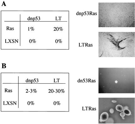
FIG. 2.
Loss of p53 is not sufficient for Ras to overcome contact inhibition or for anchorage-independent proliferation. (A) Percentage of Ras or LXSN-infected Schwann cells, expressing dnp53 or SV40 LT (LT), which proliferated to form foci in a confluent monolayer after 2 weeks, as visualized by staining with methylene blue. A representative focus of SV40 LT cells (LT) expressing Ras is shown. (B) Percentage of Schwann cells expressing dnp53 or LT, together with Ras or the empty vector LXSN, which proliferated to form colonies in soft agar. Pools of cells (4 × 10³) were seeded into soft agar. After 2 weeks the colonies were photographed and counted. Representative colonies of cells expressing Ras, together with dnp53 or LT, are shown. Assays for panels A and B were carried out in duplicate and are representative of three independent experiments.

