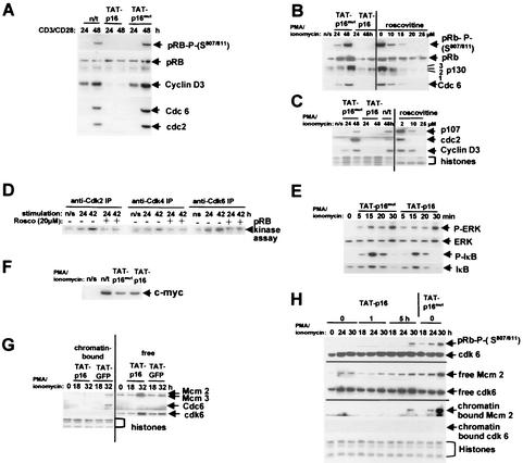
FIG. 2.
pTAT-p16INK4A and roscovitine inhibit the pRb/p130 pathways and prevent transition through the G0→G1 commitment point. (A to C) Nonactivated T cells were transduced with TAT-p16INK4A or TAT-p16MUT (inactive mutant of p16INK4A), or roscovitine was added at the concentrations shown. The cells were stimulated with CD3/CD28 (A) or PMA-ionomycin (B and C), and samples were taken at 24 and 48 h. The pRb phosphorylated on S807/811, the total pRb and p130 proteins, and the E2F-regulated Cdc6, cdc2 p107, and cyclin D3 proteins were analyzed by Western blotting. pRb, total pRb protein; 3, hyperphosphorylated p130; 1,2, hypophosphorylated p130. n/s, not stimulated with CD3/CD28 or PMA-ionomycin. n/t, not transduced with TAT-p16INK4A or TAT-p16MUT. Histones stained with Coomassie blue were a loading control for panel C. (D) Inhibition of cdk activity by roscovitine. T cells were stimulated with CD3/CD28 in the presence (+) and absence (−) of 20 μM roscovitine, and the activity of each cdk was determined by in vitro kinase assays. (E) Western blot of phospho-IκB (S32) and ERK1/2 phosphorylated at T202/Y204. Blots of total cell IκB and ERK1/2 proteins are also shown. (F) Western blot of c-myc 5 h after stimulation. n/s, not stimulated; n/t, stimulated but not transduced with TAT protein. (G) Inhibition of cdk activation prevents the formation of DNA origin recognition complexes. T cells were transduced with TAT-p16INK4A or TAT-GFP (as a control), and extracts of chromatin-bound and free proteins were prepared at the times shown. The presence of Mcm2, Mcm3, and Cdc6 in each sample was determined by Western blotting. Histones and cdk6 are loading controls for chromatin-bound and free proteins, respectively. (H) The T cells were stimulated with PMA-ionomycin and transduced with TAT-p16INK4A or TAT-p16MUT at 0, 1, or 5 h after stimulation. Samples were obtained at 18, 24, and 30 h for the analysis of pRb phosphorylation and chromatin binding of Mcm2. cdk6 was used as a loading control for total cell lysates and for extracts of non-chromatin-bound (free) protein and histones stained with Coomassie blue for chromatin. The data shown in panels B, C, and G are each representative of six separate experiments; the data shown in panels A, E, F, and H are each representative of three experiments, and the data shown in panel D are representative of two experiments.

