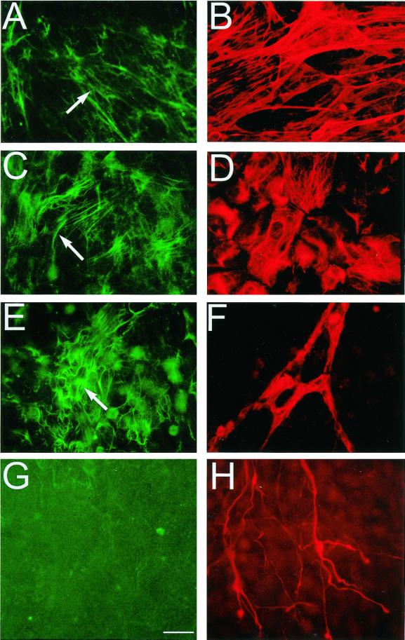
Figure 2.
Colocalization of LTBP-1 fibers with smooth muscle cells (A and B), epithelial cells (C and D), endothelial cells (E and F), and neurons (G and H). Twelve-day-old EBs were incubated with rabbit polyclonal anti-LTBP-1 serum Ab39, fixed, and incubated with either mouse monoclonal anti-smooth muscle actin antibody (B), mouse monoclonal anti-cytokeratin K18/8 antibody (D), the rat monoclonal anti-ICAM-2 antibody (F), or a mixture of two mouse monoclonal antibodies, SM31 and SM32 (H) raised against neurofilaments. Antigen–antibody complexes were detected with FITC-conjugated anti-rabbit, anti-mouse, or anti-rat IgG antibodies. Magnification, ×400. Bar, 25 μm.

