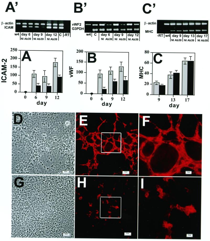
Figure 3.
Effect of anti-LTBP-1 antibody on the expression of the endothelial cell markers ICAM-2 and vWF and the formation of vessel-like structures. EBs were grown in the presence of Ab39 (1:200) or nonimmune (NI) rabbit serum (1:200) for 21 d. The expression of ICAM-2 (A′ and A), vWF (B′ and B), and αMHC (C′ and C) was measured by RT-PCR at the indicated time points. The RT-PCR products were analyzed by gel electrophoresis (Figure 3A′, B′, and C′). W4 represents samples amplified from ES cells, and C represents samples amplified from adult mouse tissue as a positive control.−RT represents samples amplified in the absence of RT. The values for the individual bands measured after densitometry of the amplification products are presented as percentages of values of the housekeeping genes β-actin or G3PDH (A, B, and C). These values represent averages from four experiments. The gray bars represent values from cells treated with nonimmune serum and the black bars represent values from cells treated with antibody 39. Nine-day-old EBs, grown in the presence of nonimmune rabbit serum (D–F) or Ab39 (G–I), were also characterized by light microscopy (D and G) and by indirect immunofluorescence (E, F, H, and I) with anti-ICAM-2 antibody to detect differentiated endothelial cells. The outlined boxes in E and H indicate the fields represented at higher magnification in F and I. Magnifications, ×200 (D, E, G, and H); ×630 (F and I). Bars, 50 μm (D and G); 10 μm (E, F, H, and I.

