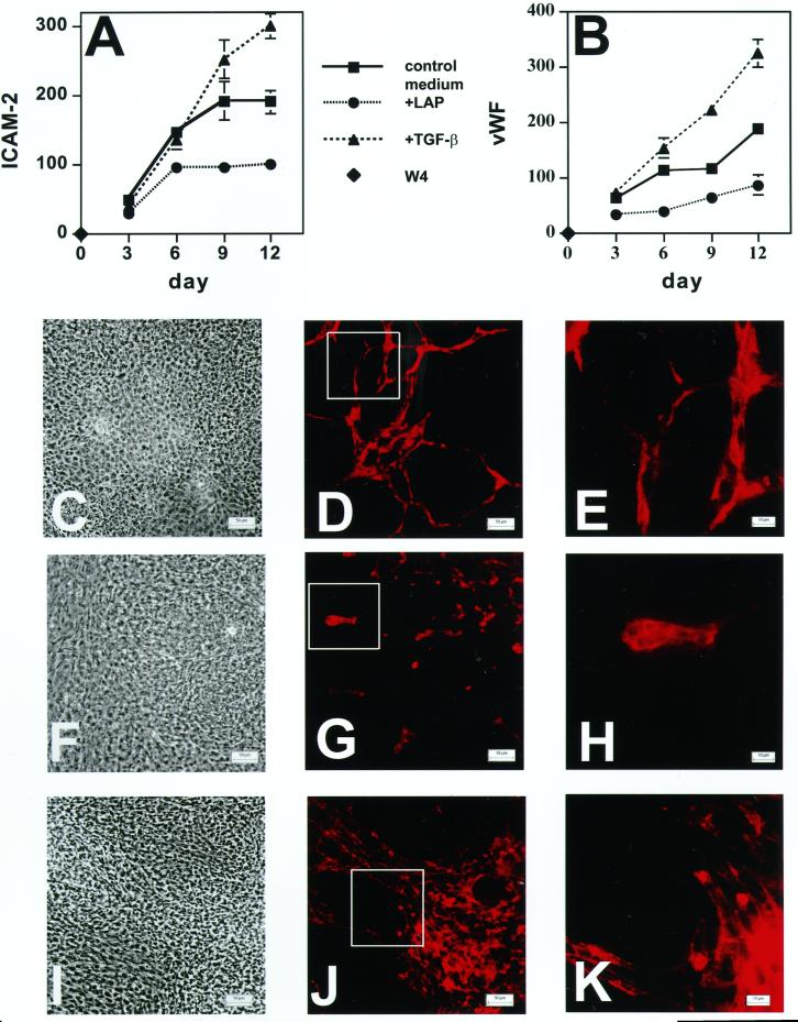
Figure 4.
Effect of LAP or TGF-β on the expression of the endothelial cell markers ICAM-2 (A) and vWF (B) and on formation of vessel-like structures in EBs (C–K). EBs were grown for 21 d in control culture medium or in medium supplemented with either recombinant LAP (3.2 μg/ml) or recombinant TGF-β1 (1 ng/ml). ICAM-2 (A) and vWF (B) expression was measured by RT-PCR. The values obtained by densitometry for the bands of each endothelial marker are presented as percentages of the values for the housekeeping gene β-actin. The values represent the averages compiled from five gels. Nine-day-old EBs grown in the presence of recombinant LAP (3.2 μg/ml; F–H), recombinant TGF-β1 (1 ng/ml; I–K), or control medium (C–E) were also characterized by light microscopy (C, F, and I) and by indirect immunofluorescence (D, E, G, H, J, and K) with anti-ICAM-2 antibody. The outlined boxes in D, G, and J indicate fields shown at higher magnification in E, H, and K, respectively. Magnifications, ×200 (C, D, F, G, I, and J); ×630 (E, H, and K). Bars, 50 μm (C, D, F, G, I, and J); 10 μm (E, H, and K).

