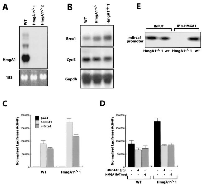
FIG. 5.
Hmga−/− cells express higher levels of BRCA1 mRNA and protein. (A) Hmga1 expression in wild-type (WT) ES cells and in two different Hmga1−/− ES cell clones analyzed by Northern blotting. (B) Northern blot showing the expression of Brca1 (top panel), cyclin E (middle panel), and GAPDH (lower panel) mRNA in WT Hmga1+/− and Hmga1−/−1 ES cell lines. (C) Activity of the human and mouse BRCA1 minimal promoter regions transfected in WT or Hmga1−/−1 ES cells. pGL3 vector was transfected as a control. Error bar, standard deviation. (D) Effect of HMGA1b overexpression on the activity of the human BRCA1 minimal promoter region transfected in WT or Hmga1−/−1 ES cells (Error bar, standard deviation.) Each transfection assay was performed in triplicate and repeated in at least three independent experiments and using two different Hmga1−/− ES cell clones. (E) ChIP assay performed on WT and Hmga1−/− (Hmga1−/−1) ES cells. The purified DNA untreated (input) or immunoprecipitated with an anti-HMGA1 antibody (IP α-HMGA1) was used as a template for the PCRs with primers that amplify the mouse Brca1 promoter region comprised between nt −236 and +44.

