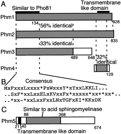
Figure 3.
(A) Homology between Phm1, Phm2, Phm3, and Phm4. Shaded and white boxes represent homologous and nonhomologous regions, respectively, and their identities to the Phm1 sequence are indicated by percentage. The region homologous to Pho81 and the putative transmemembrane region are indicated above the Phm1 box. (B) Consensus sequences in the N-terminal regions of nine S. cerevisiae proteins, including Pho81. Hydrophobic, positive- and negative-charged amino acid residues are indicated by “*,” “+,” and “-,” respectively. (C) The putative Phm5 protein structure. The region of sequence similarity to acid sphingomyelinase in human and C. elegans is indicated by a shaded box. The putative transmembrane region is indicated by a filled box.

