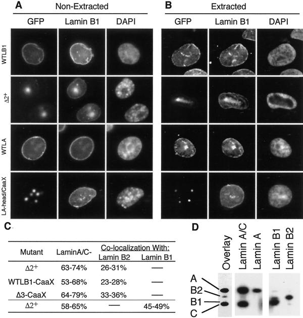
Figure 4.
Interactions between transfected mutant lamins and endogenous lamins. (A and B) CHO cells were transfected with GFP-lamins as in Figure 2. After 48 h, cells were either fixed immediately (A) or first extracted with Triton X-100 before fixation (B). Cells were then stained with antibody against CHO lamin B1 as described in MATERIALS AND METHODS. (C) Percentage of cells transfected (GFP+ cells) with the indicated mutant lamin B1 construct that either had lost detectable lamin A/C signal at the nuclear rim (Lamin A/C−) or that displayed colocalization of endogenous B-type lamins with the mutant GFP-lamins (Lamin B colocalization) was scored. Shown are the percentages obtained in two independent experiments. (D) A nuclear matrix fraction was prepared from HeLa cells and multiple aliquots of this fraction were resolved on 10% SDS-PAGE gels. Samples were transferred to nitrocellulose, the nitrocellulose was cut into separate lanes, and individual lanes were blotted with type specific anti-lamin antibodies (indicated over each lane) to reveal the positions of the lamins (indicated to the left of the overlay lane), or overlayed with GST-Δ2+ (overlay). Parallel gels, overlayed with GST-Δ1+, did not reveal any interaction of Δ1+ with any of the endogenous lamins.

