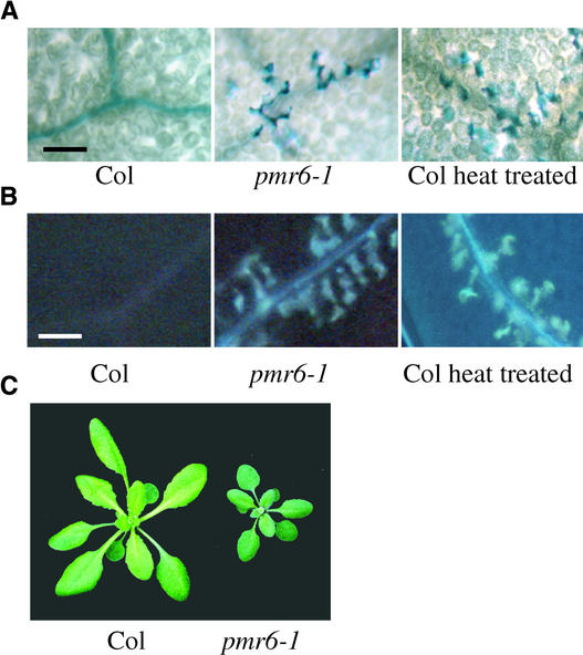
Figure 4.
Pleiotropic Effects of the pmr6 Mutation.
(A) Uninfected leaves stained with trypan blue. Dead cells stain dark blue. Individual mesophyll cells along the veins are stained in both pmr6-1 leaves and heat-treated Col leaves. Heat treatment was conducted by transferring plants to 37°C for 20 h. Leaves were stained 2 days after heat treatment. Veins appear as dark lines.
(B) Accumulation of autofluorescent compounds, as indicated by bright blue spots, follow the same pattern as dead cells in both pmr6-1 and heat-treated Col leaves. Veins appear as blue lines.
(C) Mature rosettes were photographed as they began to bolt. Note that pmr6-1 is smaller and greener than Col.
Plants were grown under a 14-h photoperiod for ∼3 weeks before being photographed. Bars in (A) and (B) = 100 μm.

