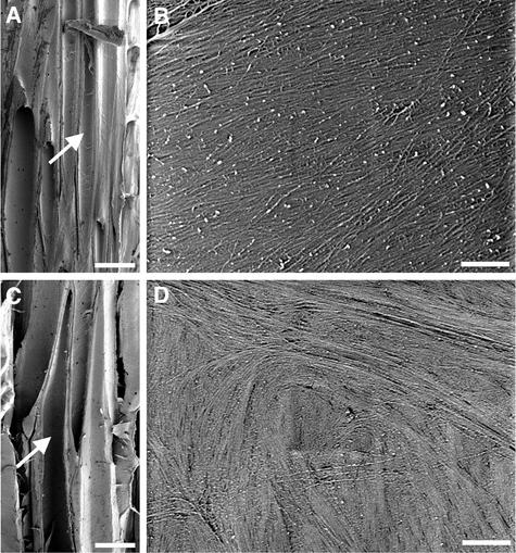
Figure 8.
Visualization of Cellulose Microfibrils in the Innermost Layer of Fiber Cell Walls.
(A) Longitudinal section of the nonelongating region of a wild-type stem showing interfascicular fiber cells (arrow).
(B) Cellulose microfibrils in the middle part of a wild-type fiber cell. Note that microfibrils run in parallel at an angle of 15 to 25° relative to the transverse orientation.
(C) Longitudinal section of the nonelongating region of a fra2 stem showing interfascicular fiber cells (arrow).
(D) Cellulose microfibrils in the middle part of a fra2 fiber cell showing a disoriented pattern.
Bars in (A) and (C) = 25 μm; bars in (B) and (D) = 0.5 μm.

