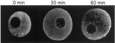
Figure 5.
Pattern of development of anti-PLC-γ1 immunostaining in whole-mouse oocytes. The selected photographs indicate the pattern of development of anti-PLC-γ1 immunostaining observed by confocal microscopy in whole-mouse oocytes analyzed at different times after release. These observations were recorded in a single optical section through the GV. No immunostaining in the nucleus at the three studied times. Diffuse PLC-γ1 immunostaining in the cytoplasm at time zero. The labeling was first intense around the plasma membrane, and then at a pole of the cortex after 30 and 60 min in culture.

