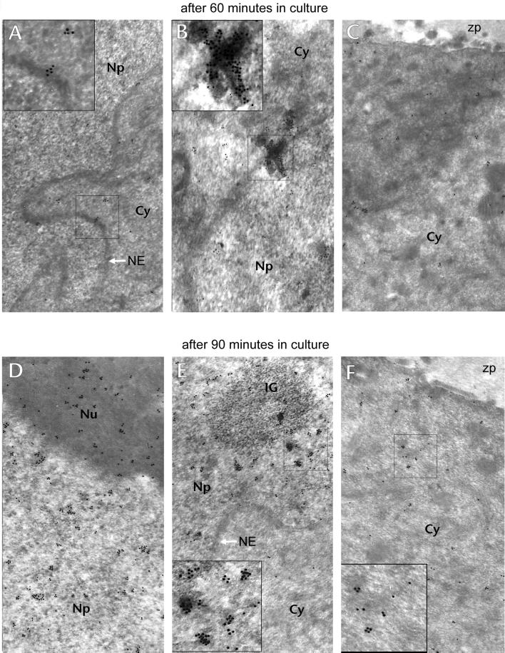
Figure 7.
Electron microscope immunocytochemistry 60 and 90 min after follicular release. (A–C) Oocytes treated with anti-PLC-β1 60 min after follicular release. (A) Gold particles are similarly distributed in the cytoplasm (Cy) and the nucleoplasm (Np); some gold particles are still present on the nuclear envelope (arrow). (B) A large aggregate of gold particles crossing the nuclear envelope. (C) Diffuse staining of the oocyte cortex and cytoplasm; zp, zona pellucida. (D–F) Oocytes treated with anti-PLC-β1 90 min after follicular release. (D) Nucleolus (Nu) is also strongly stained. (E) Gold particles are concentrated in the nucleoplasm and in the IGs; the nuclear envelope (NE) is free of particles. (F) Labeling in the cortex region is noticeable. Magnification, 40,000×. The regions selected in small squares are presented as 2× enlarged inserts.

