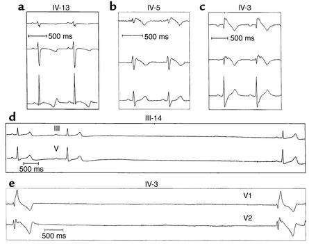
Figure 2.
EKG traces of mutation carriers showing leads V1, V2, and V5. (a) QT interval prolongation (patient IV-13 of the pedigree shown in Figure 1). (b) ST segment elevation (patient IV-5 of the pedigree). (c) ST segment elevation and right bundle branch block (patient IV-3 of the pedigree). (d) First-degree AV block and sinus arrest (patient III-14 of the pedigree).

