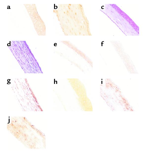
Figure 2.
TIMP-1 DNA expression and MMP-9 and neutrophil infiltration into corneas of BALB/c and MMP-9 knockout mice. BALB/c and MMP-9 knockout mice were infected with 106 PFU HSV-1 RE or UV-inactivated RE on their scarified corneas. At day 2 after infection, mice were sacrificed, and the eyes were snap-frozen in OCT compound. (a and b) TIMP-1 expression in (a) naive and (b) TIMP-1–treated groups. (c and d) Histopathology of infiltrating cells in the cornea of BALB/c. (c) Naive and (d) RE, day 2 after infection. (e–g) Immunohistochemistry for MMP-9. (e) Naive, (f) UV-inactivated RE day 2 after infection, and (g) RE day 2 after infection. (h–j) Immunohistochemistry for Gr-1. (h) UV-inactivated RE, (i) 129 Sv/Ev, and (j) MMP-9 KO. ×200.

