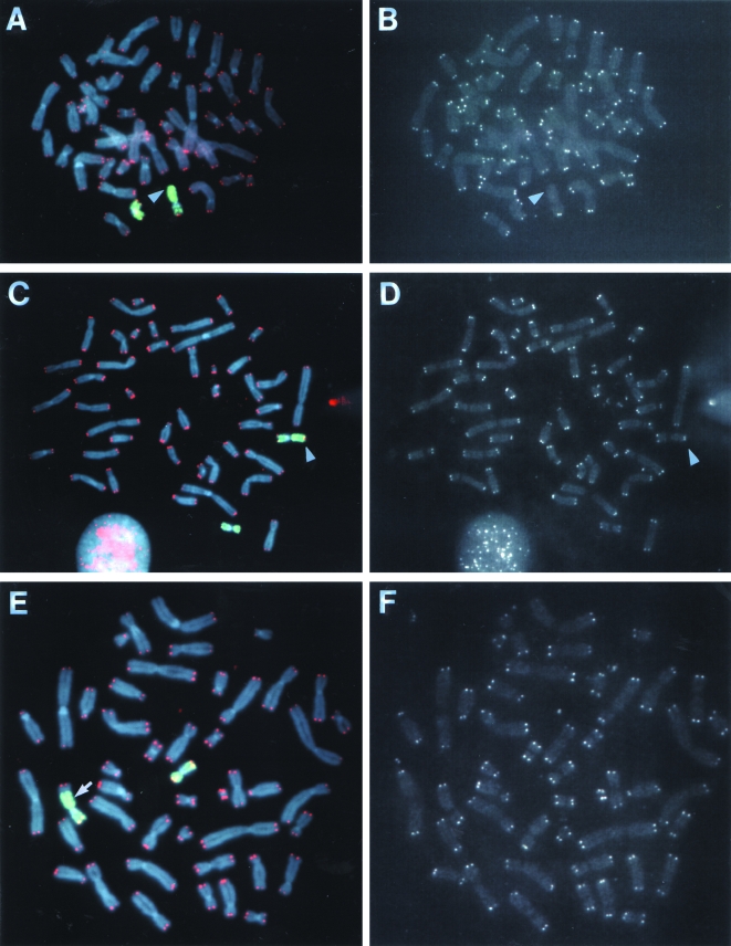
Figure 8.
Analysis of telomeres on the end of the unstable marked chromosome in subclone G71. Hybridization was performed with both a telomere-specific PNA probe (red) and a chromosome 16-specific probe (green). Chromosomes were counter stained with DAPI. Hybridization signals are shown for both probes together (A, C, and E) and with the telomere-specific PNA probe alone (B, D, and F). The results are shown for a cell (A, B) in which the DNA joined on to the end of the marked chromosome originated from chromosome 16 and had no detectable telomere on the end (arrow); a cell (C, D) in which the DNA joined on to the end of the marked chromosome originated from chromosome 16 and had a telomere on the end (arrow); and a cell (E, F) in which the DNA joined on to the end of the marked chromosome originated from both chromosome 16 and another chromosome and had a telomere on the end. The location of the interstitial plasmid sequences (as determined by hybridization with the pNCT-Δ probe, data not shown) at the junction between the marked chromosome and a fragment of chromosome 16 is shown (arrow, E).

