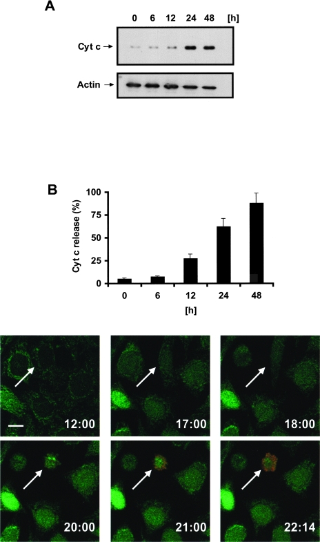
Figure 4.
Aspirin induces the release of cytochrome c from HeLa cells. HeLa-Cc-GFP cells were treated with 5 mM ASA and analyzed for cytochrome c release by means of (A) Western blot with cytochrome c antibody, (B) FACS analysis (CLAMI assay), or (C) multicolor time-lapse confocal microscopy. Cytochrome c-GFP (green) moves from a mitochondrial staining pattern to a cytoplasmic distribution. Loss of plasma membrane integrity was detected by uptake of propidium iodide (red). The arrow indicates a cytochrome c-releasing cell; the number at the bottom right of each frame represents the time after treatment with aspirin. Images were taken with a x60 objective and zoomed 1.8 times. Scale bar= 15 µm.

