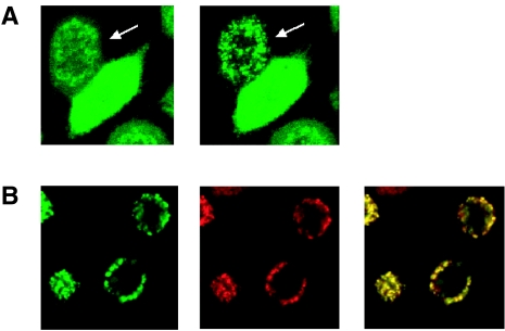
Figure 8.
Aspirin induces caspase-independent translocation of Bax to mitochondria. HeLa-Bax-GFP cells were treated with 5 mM aspirin and Bax-GFP was monitored by time-lapse confocal microscopy. (A) After treatment with aspirin Bax-GFP changed from a diffuse cytoplasmic to a perinuclear pattern in the presence or absence (data not shown) of zVAD-fmk (100 µM). The arrow indicates a cell with Bax translocation to the mitochondria. Images were taken with a x40 objective and zoomed 2.0 times. (B) Left image (green) is translocated Bax-GFP after aspirin treatment. Middle image (red) is the mitochondrial marker TMRE (40 nM). The majority of translocated Bax-GFP is colocalized with TMRE as seen by overlaying the green and red images (yellow).

