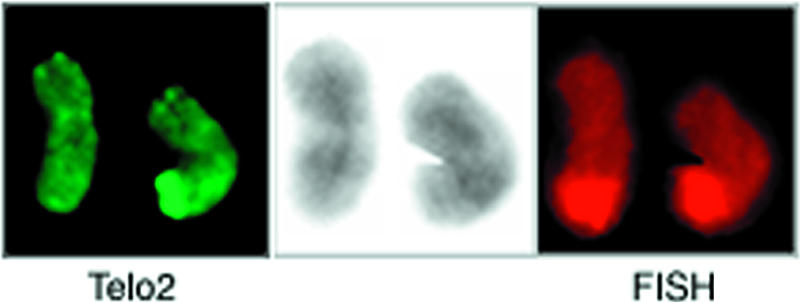
Figure 4.

Staining of the 11q terminus by PRINS painting in a pair of chromosomes 11, one of which was positive for the 11q telomere expansion. The telomeric repeats were stained with Telo2 (left), whereas the tip of 11q was painted by FISH (right). The DAPI staining is shown at the center. The two 11q termini appeared equally stained by the FISH probe, and no site outside 11q showed apparent staining.
