Figure 1.
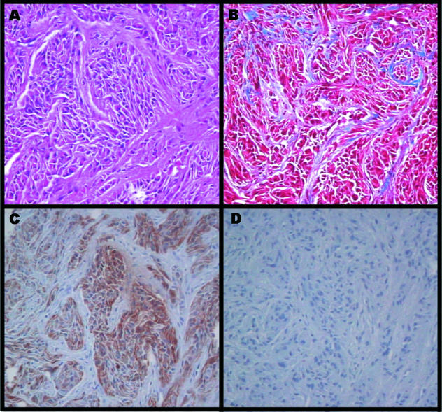
Histological view of subcutaneous tumor generated from subcutaneous allogeneic astrocytoma implant into fetal dogs. H&E stained section (A) shows bundles of fibrillary malignant cells within a network of connective tissue. Trichrome staining (B) shows a rich collagenous network of fibers around the tumor cell bundles. Immunostaining with anti-GFAP antibodies (DAKO Corp., Carpinteria, CA). (C) portrays a strong presence of this glial-specific intermediate filament protein. (D) The isotype matched negative section. (Original magnification, 40 x objective).

