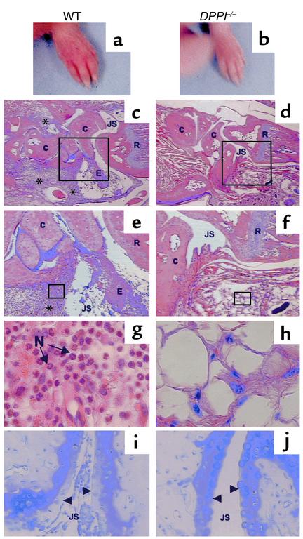
Figure 3.
Clinical and histological assessment of monoclonal antibody–induced arthritis. Forepaw of a WT mouse (a) and a DPPI–/– mouse (b) on day 7 after receiving arthrogenic antibodies against type II collagen. Note the redness and swelling involving the entire paw, including the digits, of the WT mouse (a). Histological analysis of sections taken from the radiocarpal joints of immunized animals, stained with hematoxylin and eosin (c–h), and toluidine blue (i and j) (to detect proteoglycan). WT mice (c, e, g, and i) have marked leukocyte infiltration (*) in the subsynovial tissue and joint space, adherence of inflammatory cells to joint surfaces, and proteoglycan loss as evidenced by the reduction in metachromasia at the superficial border of the cartilage matrix (arrowheads). The infiltrating cells are predominantly neutrophils (N) (g). DPPI–/– mice were protected from arthritis, as evidenced by normal joint histology, lack of inflammatory infiltrates, and preservation of proteoglycan content (d, f, h, and j). Magnification: c and d, ×16; e and f, ×40; g and h, ×400. E, exudate; C, carpal bones; R, radius; JS, joint space.

